Free animal cell coloring pages Generator & Download PNG or PDF
Just enter text to create your own custom animal cell coloring pages in minutes – Download in PNG or PDF for printing and coloring fun!
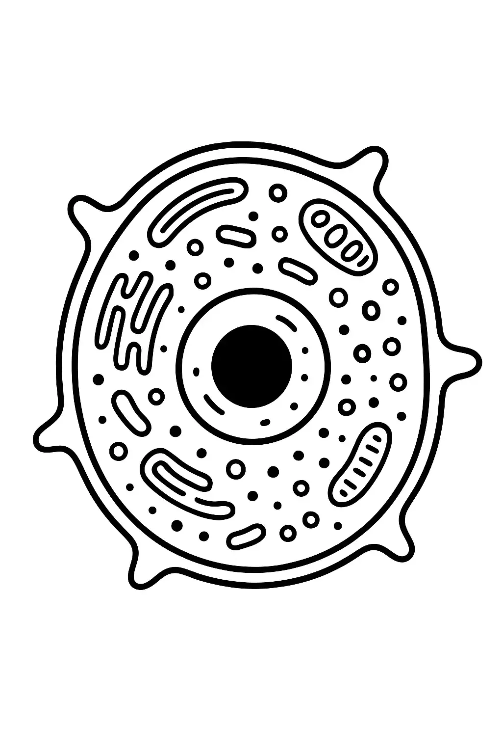
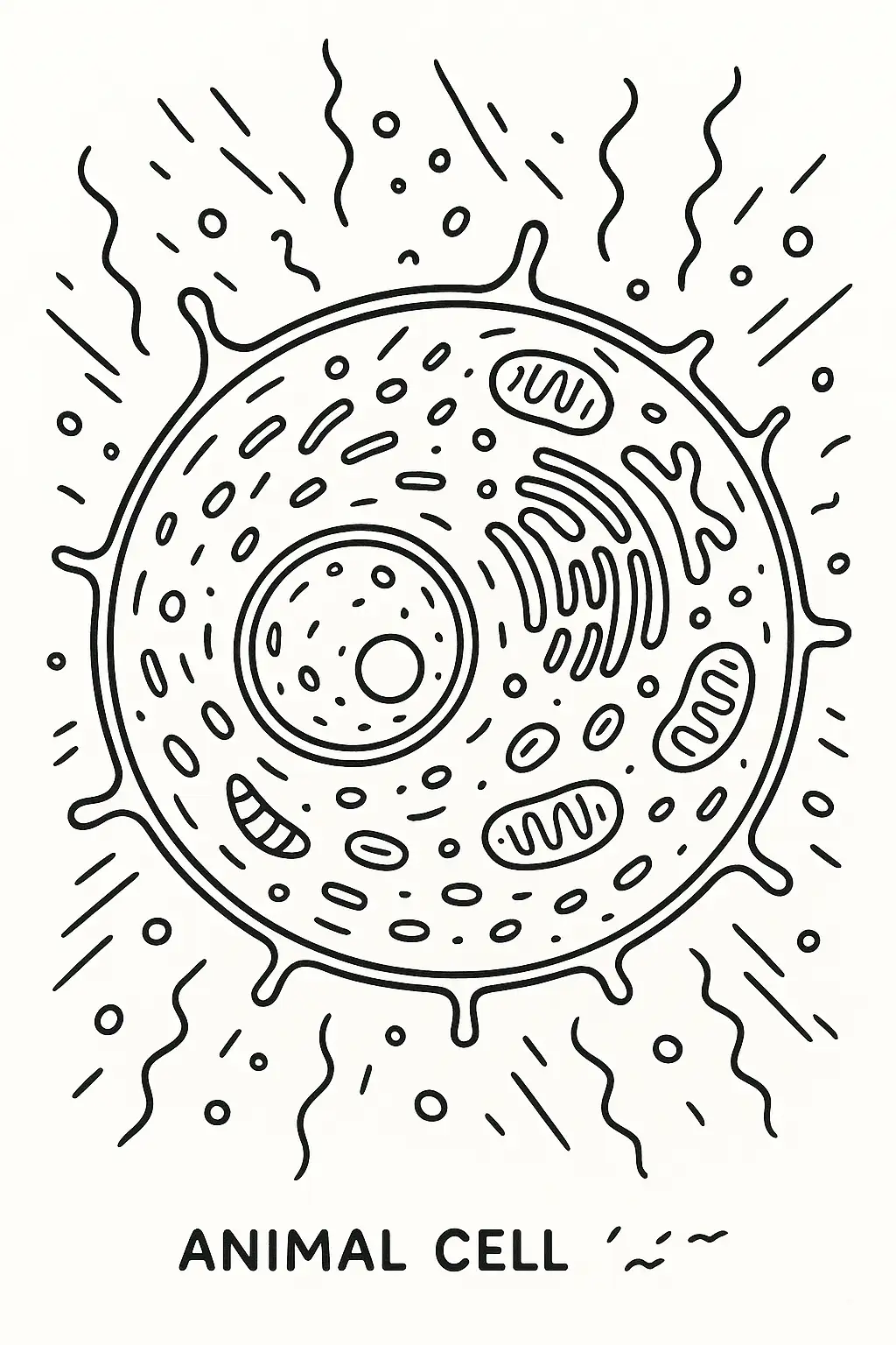
A coloring page of an animal cell with a clearly - defined round nucleus in the center. The nucleus has a dark - colored nucleolus inside. Surrounding the nucleus, draw the cytoplasm as a light - colored, jelly - like substance. The cell membrane should be a thin, dark line enclosing the entire cell.
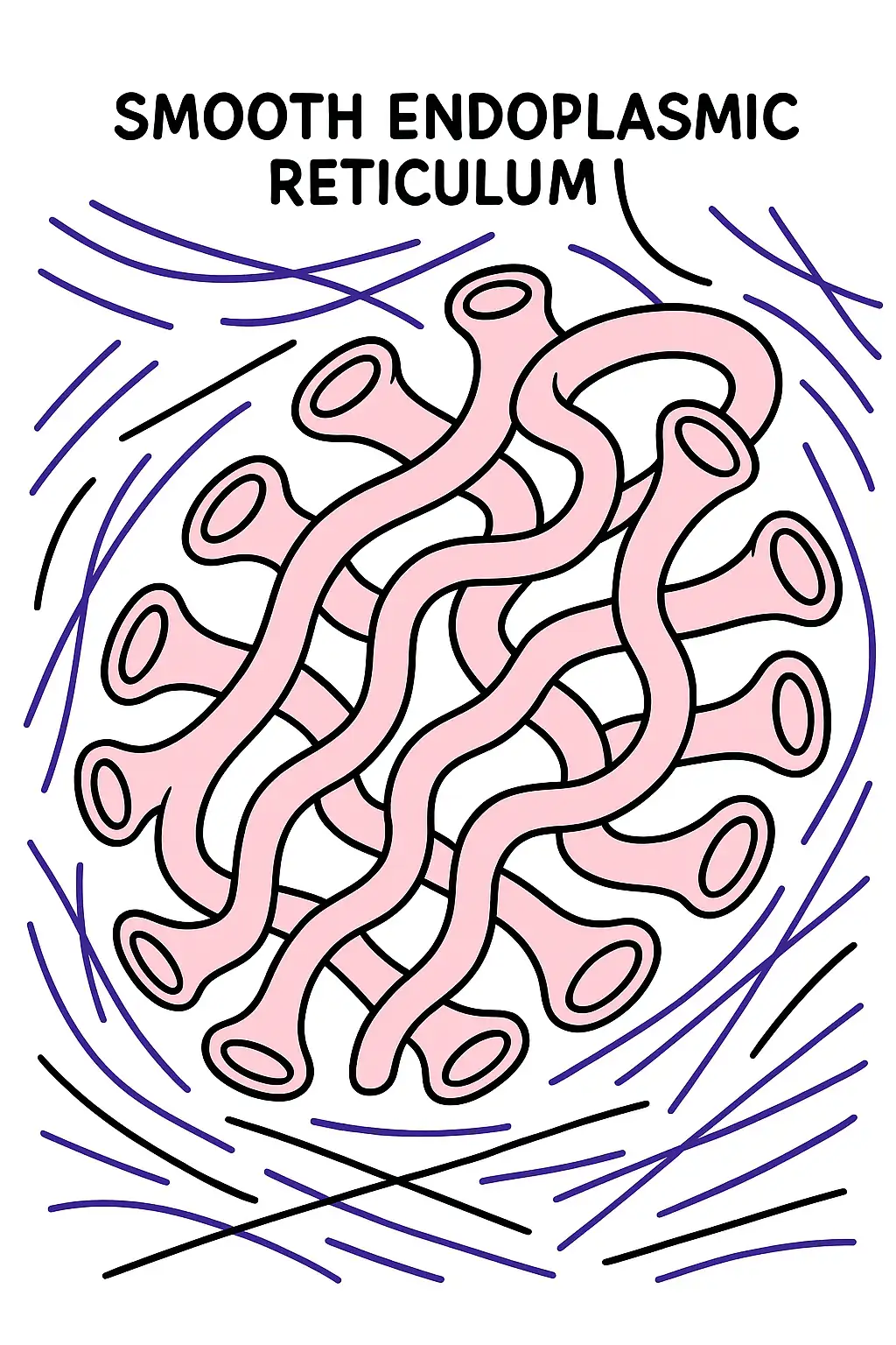
Generate a coloring page where the smooth endoplasmic reticulum is shown as a network of tubes. It has a different appearance from the rough endoplasmic reticulum as it lacks ribosomes. Color it a pale pink. In the cytoplasm, also include some microfilaments and microtubules, thin and thick lines respectively, colored purple.
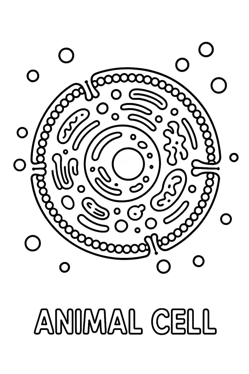
Design an animal cell coloring page where the cell is interacting with its environment. There are some molecules (small circles of different colors) outside the cell, and some are being transported through the cell membrane. The cell membrane should be colored in a way to show its semi - permeable nature, perhaps with a gradient of colors.
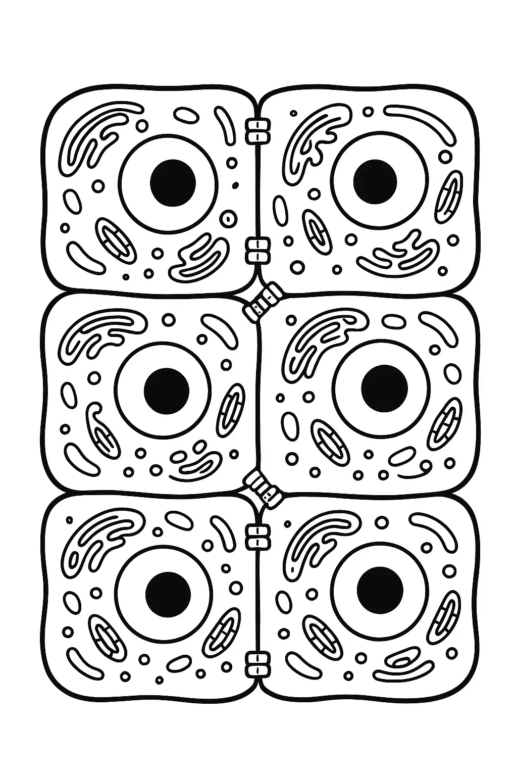
Sketch a coloring page where the animal cell is part of a tissue. Draw a few adjacent cells with their cell membranes touching. You can show some cell - to - cell junctions, like tight junctions or gap junctions, colored yellow between the cells.

Generate a coloring page where the animal cell is repairing itself. Some vesicles are moving towards a damaged part of the cell membrane, and the cytoplasm has some proteins and enzymes (small, irregular - shaped molecules) working on the repair process. Color the vesicles yellow and the repair - related molecules purple.
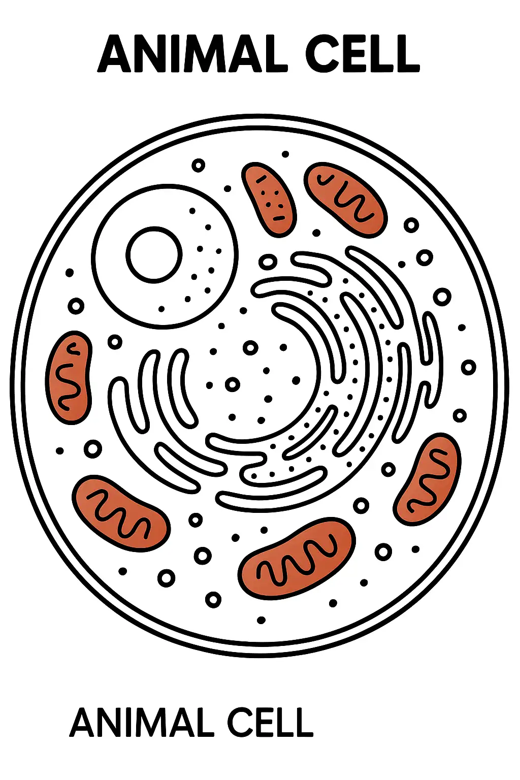
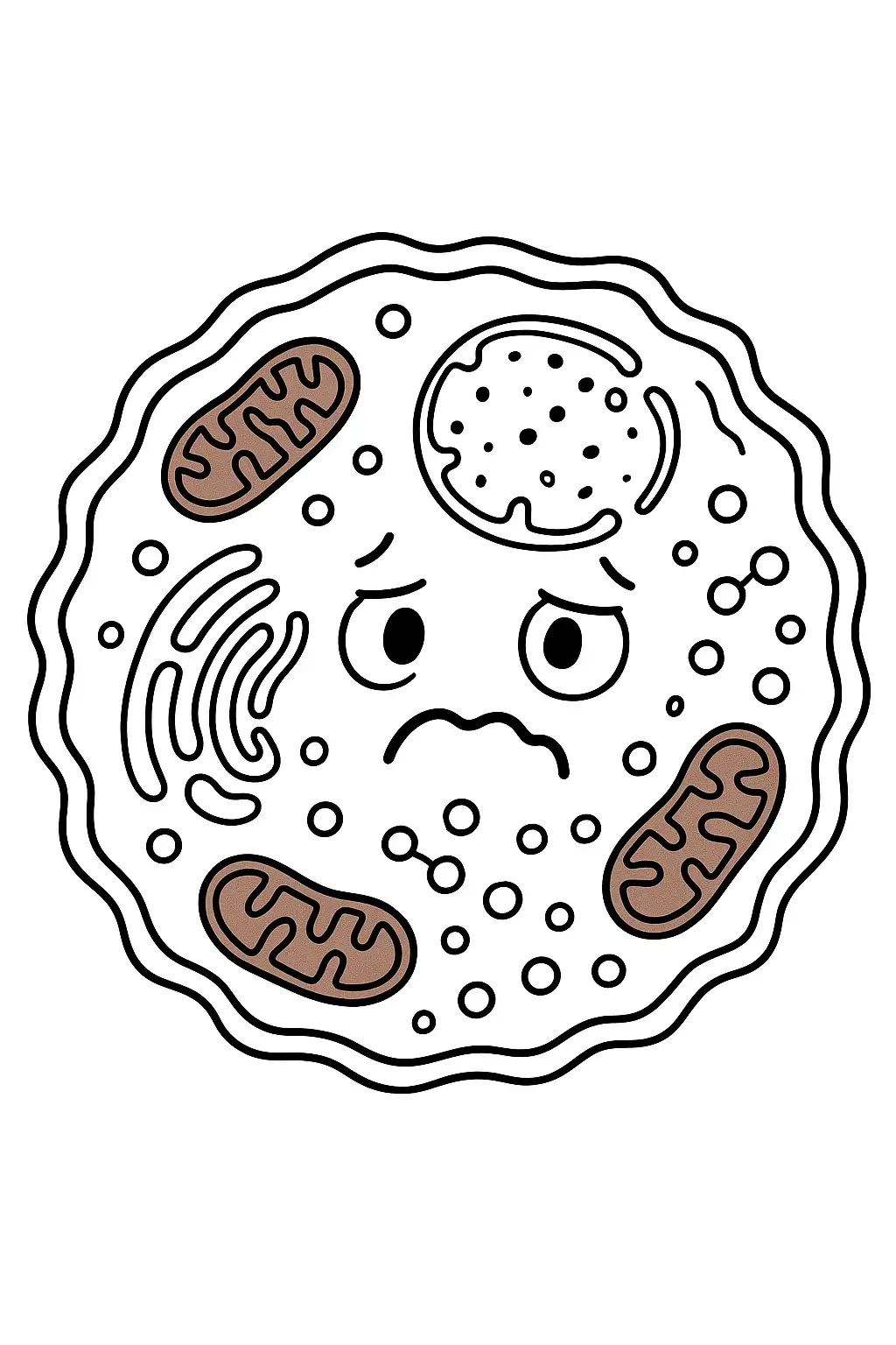
Create an animal cell coloring page where the mitochondria are shown as small, bean - shaped structures scattered throughout the cytoplasm. Color the mitochondria a reddish - brown to represent their energy - producing function. Add some ribosomes, small dots, on the rough endoplasmic reticulum, which looks like a series of flattened sacs.
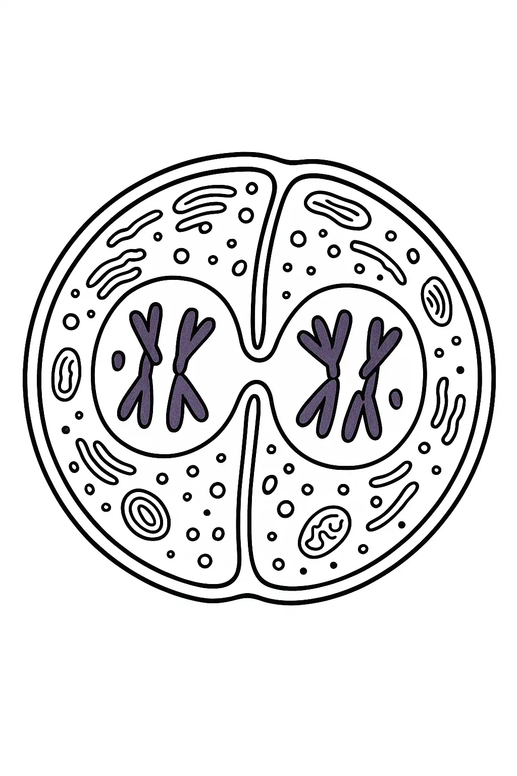
Make an animal cell coloring page with a cell that is in the process of dividing. Show the nucleus undergoing mitosis, with the chromosomes (colored dark purple) visible and separating. The cell membrane is starting to pinch in the middle, indicating cytokinesis.
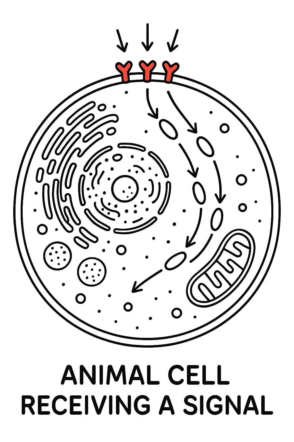
Draw a coloring page with an animal cell that has just received a signal. The signal - receiving receptors on the cell membrane (small, irregular - shaped structures) can be colored red. Inside the cell, show the initial steps of the signal transduction pathway, with some molecules starting to move in response.
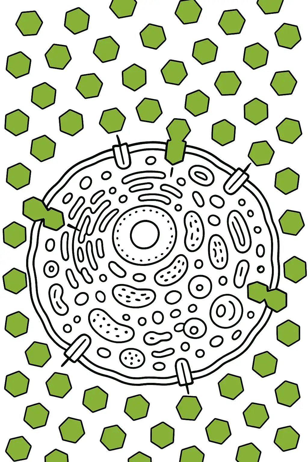
Create a coloring page of an animal cell in a nutrient - rich environment. There are many glucose molecules (hexagon - shaped) outside the cell, and some are being transported into the cell through specific transporters on the cell membrane. Color the glucose molecules green.
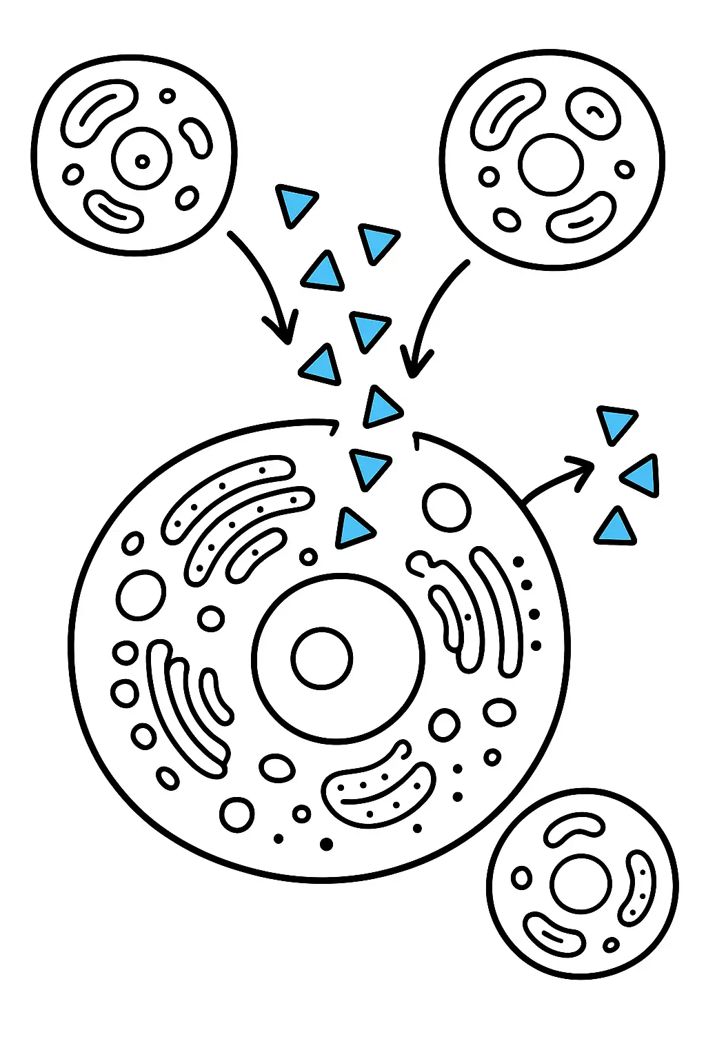
Make an animal cell coloring page with a cell that is communicating with other cells through chemical signals. There are small signaling molecules (triangular - shaped) being released from the cell and traveling towards neighboring cells. Color the signaling molecules blue.
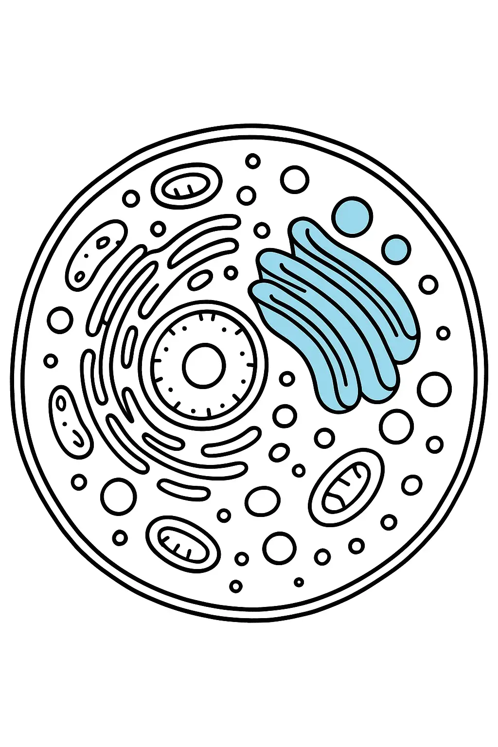
Design a coloring page featuring an animal cell with a large Golgi apparatus. The Golgi apparatus should be drawn as a stack of curved, flattened membranes. Color it a light blue. Next to it, draw vesicles, small circular structures, which can be colored yellow as they transport materials in and out of the Golgi.
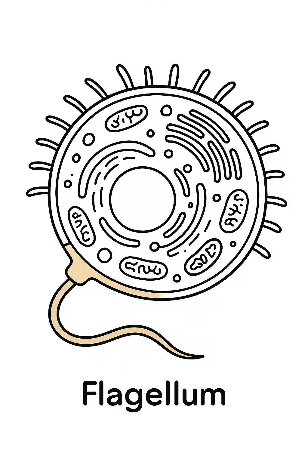
Sketch a coloring page where the animal cell has a cilia or flagella. If it's cilia, draw short, hair - like projections on the cell surface; if it's flagella, draw a long, whip - like tail. Color them a light brown. The cell membrane around the area of the cilia or flagella should be highlighted.
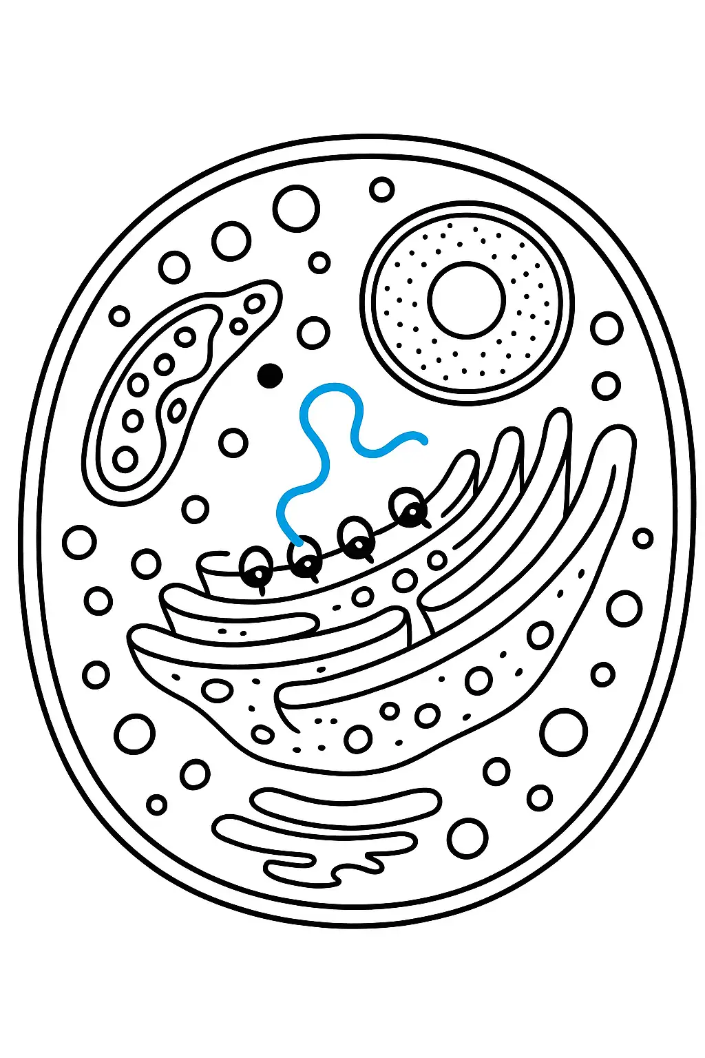
Generate a coloring page where the animal cell is producing a protein. The ribosomes on the rough endoplasmic reticulum are actively synthesizing the protein. Color the ribosomes black and the protein chain (a long, winding line) a bright blue as it emerges from the ribosome.
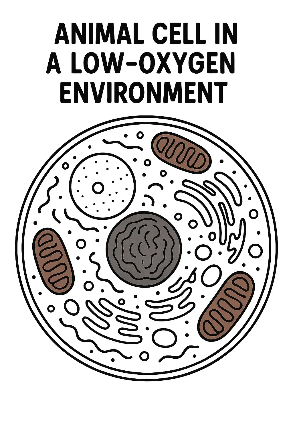
Design an animal cell coloring page where the cell is in a low - oxygen environment. The mitochondria are working harder, so color them a deeper red - brown. There may also be some changes in the cytoplasm's appearance, perhaps a darker shade to represent the cell's adaptation.
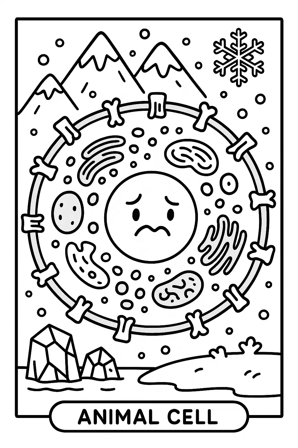
Sketch a coloring page where the animal cell is in a cold environment. The cell membrane may be changing its fluidity, and some of the proteins in the membrane are adjusting their conformation. Color the cell membrane a slightly different shade to represent these changes.
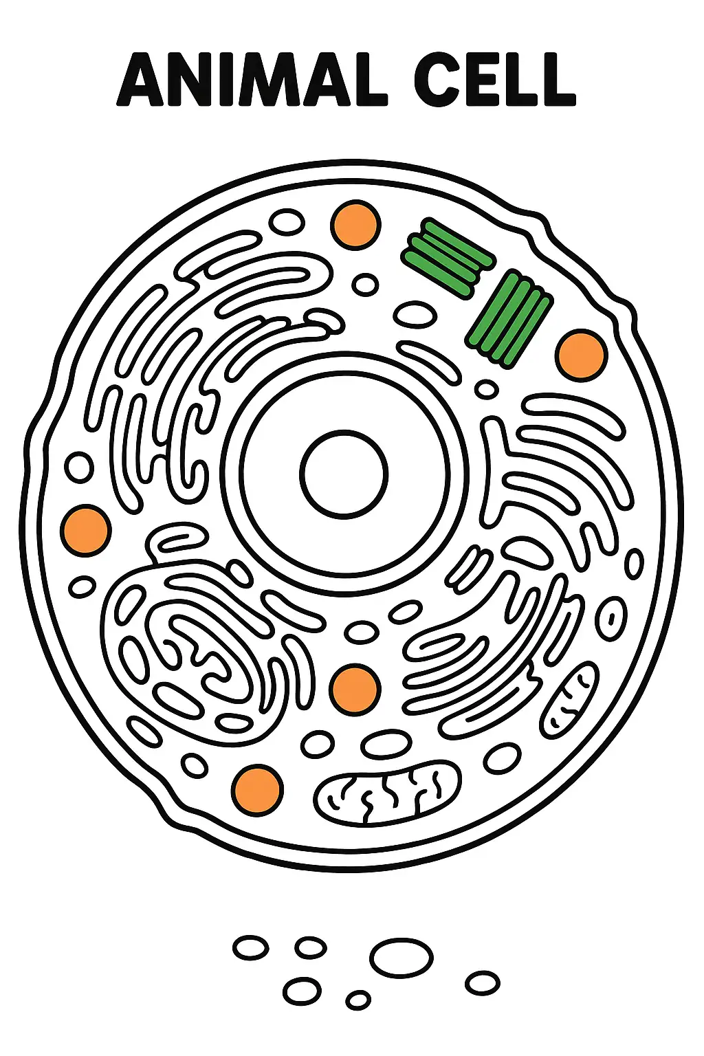
Draw an animal cell coloring page with lysosomes. These are small, spherical organelles. Color them a bright orange to signify their role in digestion. The centrioles, two small, cylindrical structures near the nucleus, can be colored green.
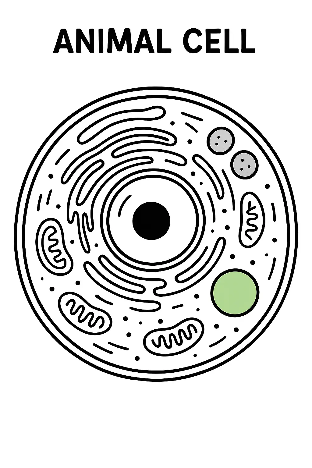
Create a coloring page of an animal cell with a vacuole. Although animal cells usually have smaller vacuoles compared to plant cells, draw a small, circular vacuole in the cytoplasm and color it light green. Next to it, draw peroxisomes, small, round organelles colored gray.
Make an animal cell coloring page with a cell that is under stress. Some of the mitochondria may be showing signs of damage, perhaps a different shade of red - brown. The cell membrane may also look a bit distorted, and there could be some free - floating molecules in the cytoplasm that are a result of the stress response.
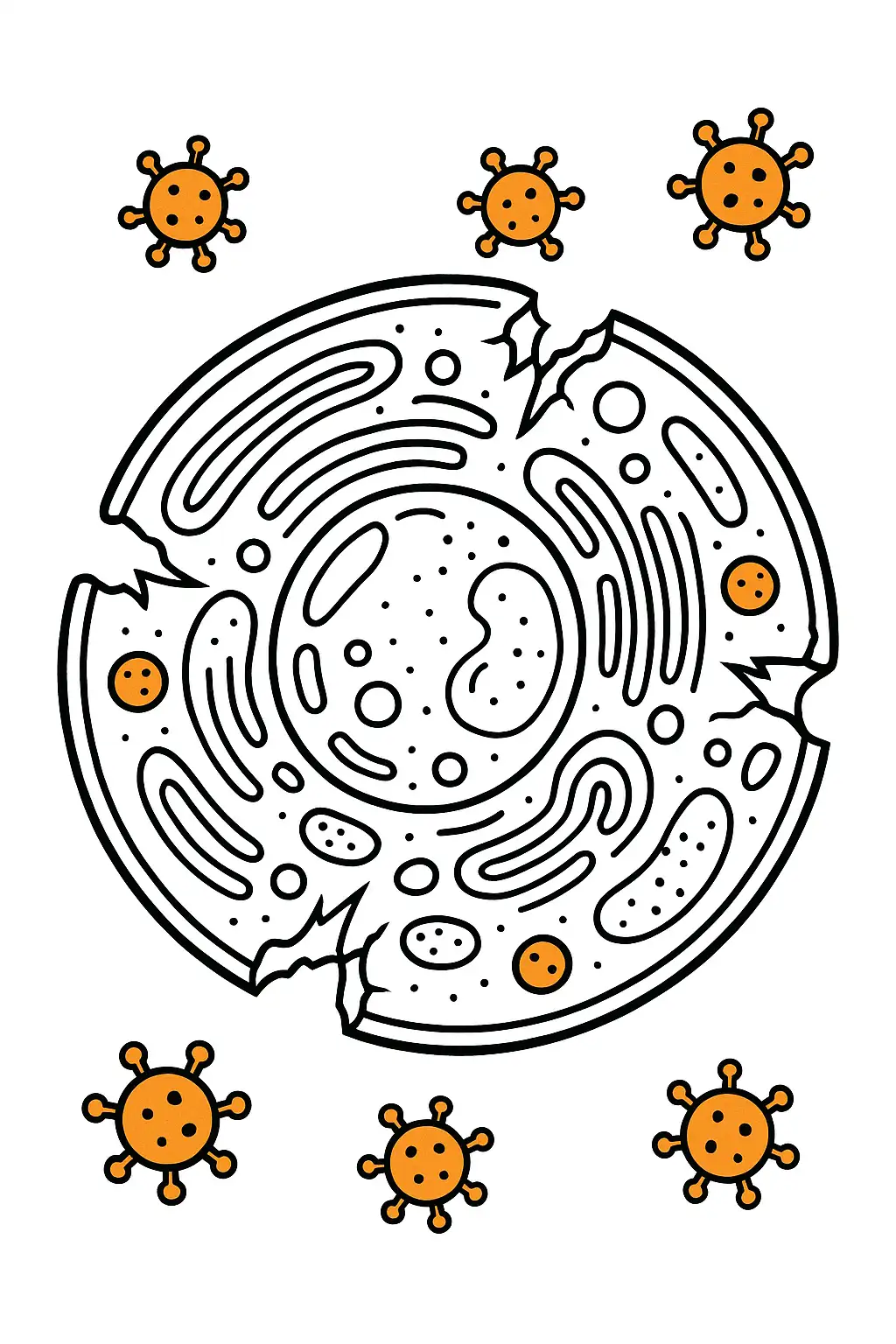
Draw a coloring page with an animal cell that is infected with a virus. Show the virus particles (small, round or hexagonal structures) inside the cell. The cell may be starting to show signs of damage, like a broken cell membrane in some areas. Color the virus particles a different color, like orange, to distinguish them.
Create a coloring page of an animal cell in a warm environment. The cell is more active, with more movement of molecules in the cytoplasm. Show some of the molecules moving around in a more energetic way, and color the cytoplasm a lighter, more vibrant color.
Related Tags
Animal cell coloring pages are a fun and interactive way to explore biology while enhancing creativity. With GenColor.ai, you can instantly generate detailed diagrams of animal cells that can be printed and colored. These coloring sheets not only spark imagination but also help students, teachers, and homeschooling parents learn the structure of an animal cell in a hands-on way. By labeling and coloring the cell parts, learners can better understand essential components such as the nucleus, mitochondria, and cytoplasm.
Learn Biology with Animal Cell Coloring Pages
GenColor.ai makes it simple to generate and download free animal cell coloring pages without needing to sign up. You can type a text prompt like 'simple animal cell diagram for coloring' or upload your own reference image, and the AI will convert it into a clean, printable coloring sheet. All downloads are available in high-resolution PNG and PDF formats, making them perfect for classrooms, science projects, or home activities.
Create Free Printable Animal Cell Coloring Pages with GenColor.ai
Each coloring page can be personalized with GenColor.ai’s editor. From realistic scientific diagrams to simplified outlines for younger learners, you can adjust the style of the coloring page by editing the prompt. This makes it easy to create different variations of animal cell diagrams for different grade levels, whether you want a highly detailed sheet for advanced biology students or a simplified version for elementary learners.
Coloring animal cells helps children and students engage more deeply with science by combining creativity and learning. Teachers can use these coloring sheets as part of lesson plans, while students can reinforce their knowledge through hands-on practice. With AI-generated coloring pages, you can always ensure variety, clarity, and accuracy in the cell diagrams you provide for learning.
Educational Benefits of Animal Cell Coloring Pages